͏‌ ͏‌ ͏‌ ͏‌ ͏‌ ͏‌ ͏‌ ͏‌ ͏‌ ͏‌ ͏‌ ͏‌ ͏‌ ͏‌ ͏‌ ͏‌ ͏‌ ͏‌ ͏‌ ͏‌ ͏‌ ͏‌ ͏‌ ͏‌ ͏‌ ͏‌ ͏‌ ͏‌ ͏‌ ͏‌ ͏‌ ͏‌ ͏‌ ͏‌ ͏‌ ͏‌ ͏‌ ͏‌ ͏‌ ͏‌ ͏‌ ͏‌ ͏‌ ͏‌ ͏‌ ͏‌ ͏‌ ͏‌ ͏‌ ͏‌ ͏‌ ͏‌ ͏‌ ͏‌ ͏‌ ͏‌ ͏‌ ͏‌ ͏‌ ͏‌ ͏‌ ͏‌ ͏‌ ͏‌ ͏‌ ͏‌ ͏‌ ͏‌ ͏‌ ͏‌ ͏‌ ͏‌ ͏‌ ͏‌ ͏‌ ͏‌ 
Is this email not displaying correctly? View it in your browser.
Image description

Infection of Neospora caninum in Dogs

Neospora caninum is a type of protozoan parasite that is closely related and bears several similarities to Toxoplasma gondii. This parasite is responsible for causing abortion in cattle worldwide and also leads to neurological diseases in young calves, dogs, and other mammalian hosts. It is known that domestic dogs and other canids act as definitive hosts, while cattle serve as the major intermediate hosts.

Life Cycle and Transmission

The life cycle of N. caninum is characterized by three infectious stages: tachyzoites, tissue cysts, and oocysts. Tachyzoites and tissue cysts, which contain bradyzoites, are found in the intermediate and definitive hosts and occur intracellularly. Domestic dogs and other canids are the only definitive hosts for N. caninum, with sporulation of oocysts occurring outside the host.

Neospora caninum
is transmitted both transplacentally and postnatally. Postnatal transmission can occur through ingestion of tissues infected with tachyzoites or tissue cysts, or through ingestion of food or water contaminated with sporulated oocysts.

Image description

Clinical signs

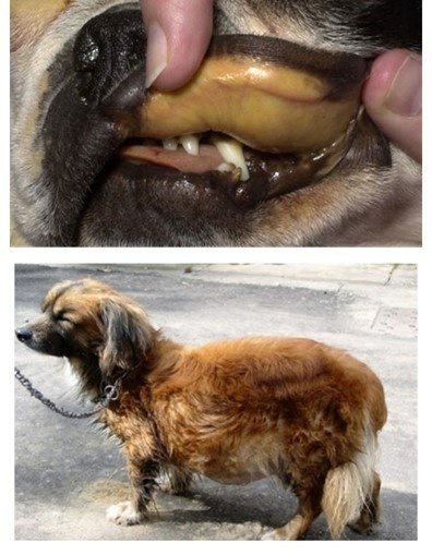
Dogs usually don't exhibit signs of infections. However, some puppies and adult dogs may show signs of infection. Puppies that get infected before or shortly after birth are often severely affected and might not survive. Symptoms in puppies usually involve nerve and muscle abnormalities. Young puppies often experience partial paralysis of their legs, particularly the hind legs. In adult dogs, neurological signs, skin inflammation with sores, liver inflammation, pneumonia, and heart inflammation may occur.

Young dogs (under 6 months old) experience more frequent and severe infections. Clinical signs are similar to toxoplasmosis, with more pronounced neurological and muscular abnormalities.

Diagnosis

Most animals infected with N. caninum show no clinical signs, so laboratory tests are required for diagnosis. Commonly used tests include:

  • Histopathology and immunohistochemistry- highly recommended in cases of reproductive problems such as abortion and fetal lesions
  • Serology tests- indirect fluorescent antibody test (IFAT), Neospora-agglutination test, and ELISA for detection of antibody levels
  • Molecular diagnosis- real-time PCR to detect the organisms or differentiate them from other protozoa

Treatment

Neosporosis treatment is usually challenging and may only provide temporary, partial, or no relief. It often requires extended periods of treatment. Dogs with neurological symptoms typically have a poor prognosis, and treatment is most effective in the early stages before muscular contracture sets in.

Most protocols for treating neosporosis aim to control clinical manifestations rather than achieve parasitological cure. Popular protocols include:

  • Clindamycin (7.5–15 mg/kg, PO or SC, q8h) for 4–8 weeks
  • Trimethoprim–sulfonamide (15–20 mg/kg, PO, q12h) for 4–8 weeks
  • Pyrimethamine–sulfonamide (5–30 mg/kg, PO, q12 h) for 4–8 weeks.
  • Ponazuril (20 mg/kg, PO, q24h) for 4 weeks

Note

Bioguard’s Qmini PCR can detect Neospora caninum DNA in 90 minutes at your clinics using feces or EDTA-blood as samples.

To learn more about Qmini PCR, click here

Want to stay up-to-date with what is going on?

Image description
Image description
Image description

Follow our Pages for the latest updates

Canine Chronic Hepatitis

Chronic hepatitis is a condition in dogs that may occur due to various disease processes. It indicates a previous occurrence of inflammation and possibly cell death in the liver. The inflammation is caused by the infiltration of different types of white blood cells that are involved in the immune system. Necrosis, which refers to the death of a large number of liver cells, may also occur.


The invasion of white blood cells and cell death in a dog's liver can be due to previous damage caused by infectious agents like viruses or bacteria or as a result of toxic damage. Toxic damage may occur due to poisons ingested by the dog or abnormal accumulation of substances required by the body, such as copper. Though some breeds like Bedlington Terriers, Labrador Retrievers, Doberman Pinschers, Dalmatians, West Highland White Terriers, Welsh Corgis, Keeshonds, and others are notably affected, this toxicity can affect any breed of dog. Copper accumulation in hepatocytes can cause oxidative damage, and its presence can escalate hepatocyte damage caused by other factors. Until dietary recommendations are modified and implemented, this problem will continue to affect canines. Inflammation and cell death can also occur due to a primary attack by the immune system against liver cells, known as an "autoimmune" disease.

Chronic vs Acute


The term "chronic" refers to a condition that has been causing damage for a prolonged period, typically lasting several weeks or more. On the other hand, "acute" hepatitis is usually characterized by a shorter duration of just a few days. While some cases of acute hepatitis can be treated successfully, many types of chronic hepatitis are not curable. However, with appropriate treatment and close monitoring, a significant number of patients with chronic hepatitis can maintain a good quality of life with minimal clinical symptoms for an extended period.

Chronic hepatitis can affect any breed of dog regardless of gender or age, although it is more common in middle-aged or older dogs. Certain breeds may be more prone to specific types of hepatitis. For instance, some breeds may develop chronic hepatitis due to the accumulation of copper in the liver cells. The excessive concentration of copper harms the liver cells and, if left untreated, typically leads to severe chronic hepatitis.

Symptoms

The liver has multiple functions, therefore the clinical signs associated with liver disease can vary significantly.

Symptoms of hepatitis in dogs can include:
• Lack of appetite
• Lethargy
• Vomiting
• Increased urination
• Excessive thirst and urination
• Swollen belly filled with fluid (ascites)
• Yellowish gums (jaundiced) and moist               tissues
• Seizures, mental dullness

Image description

Diagnosis

Apart from obtaining a detailed history of the affected dog's health prior to the appearance of symptoms, the next step would be to conduct a comprehensive physical examination of the dog, which includes carrying out a blood chemical profile, a complete blood count, an electrolyte panel, and a urinalysis. The results of the bloodwork will enable your veterinarian to identify any signs of impaired kidney function.

To further evaluate the disease, complete abdominal ultrasonography is essential. This diagnostic tool screens for concurrent diseases and helps acquire bile. It's important to note that the liver can appear normal upon examination, and changes seen in chronic hepatitis can include uniform increases in liver echogenicity, decreased distinction of portal vein margins, and normal to small liver size. Abnormalities in the liver parenchyma, biliary tree, portal vein, and peritoneum should be assessed, along with acquired shunting and the presence of free peritoneal fluid.

In order to accurately diagnose chronic hepatitis, a liver biopsy is necessary. There are 3 ways to perform a liver biopsy, surgically, via laparoscopy, or through the skin using a special needle under ultrasound guidance. However, surgical or laparoscopic biopsies are more informative compared to ultrasound-guided biopsies. The information obtained from the biopsy is required to determine the type and severity of liver disease, as well as to assess your dog's prognosis.

Treatment


The treatment for chronic hepatitis in dogs is a complex process that depends on the severity and type of liver disease, as well as the clinical signs exhibited by the dog. In severe cases, hospitalization, intravenous fluid therapy, and supportive care may be required.

The most common medications prescribed for treating the disease are immunosuppressive or anti-inflammatory. In some cases, dietary changes may be necessary. Certain medications may also be used in specific situations, such as dogs whose illness is linked to copper accumulation, abdominal fluid build-up, or neurological symptoms.

Life Expectancy


Although this condition cannot be cured with the available treatments, the good news is that the dog can still live a good quality of life for months and even years with continued therapy. Regular blood work is necessary to ensure the dog is responding well to the treatment. This helps to adjust the medication and keep the dog relatively free of clinical signs.

Image description

For any direct inquiries, please contact us at : [email protected].

About Bioguard Corporation

The Bioguard is a company focusing on animal disease diagnostic services and products.
Our animal health diagnostic center is the first and only ISO/ IEC 17025 accredited animal disease testing laboratory in Taiwan and China.


Copyright © Bioguard Corp., All rights reserved.


Our mailing address is: [email protected]

If you want to unsubscribe, click here.