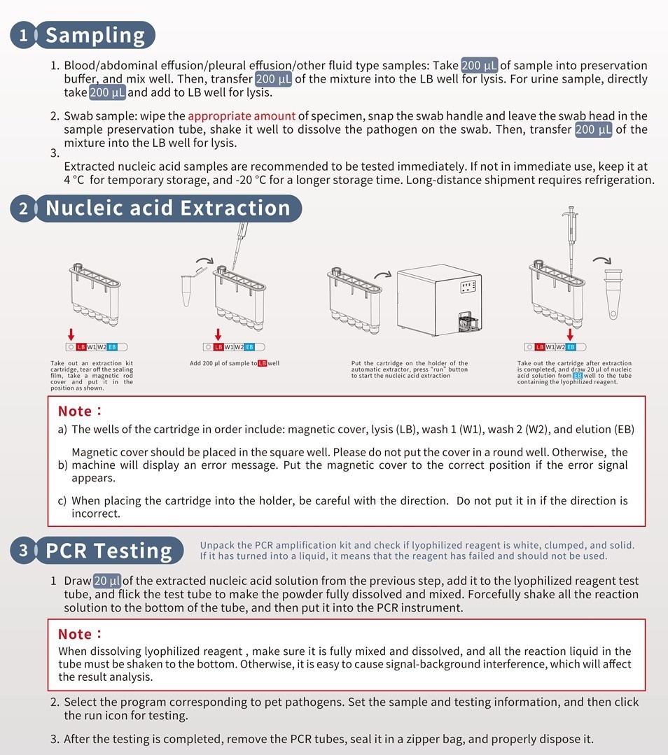
Tritrichomonas foetus is a significant cause of large intestinal diarrhea and chronic colitis in cats, according to various studies (Gookin 1999, Gookin 2001, Levy, 2003, Bell 2010). Trichomonads are flagellated protozoa with a pear-shaped body and an undulating membrane. They are similar to Giardia in size but do not have a cyst stage and are directly transmitted between hosts as trophozoites. Trichomonads thrive in moist, warm, and anaerobic conditions. T. foetus causes venereal infections in cattle and colonizes the colon and distal ileum in cats. The infection rate of T. foetus is highest in densely housed young cats in catteries and shelters. A study on purebred show cats revealed that 31% of 117 cats from 89 catteries were infected with the parasite (Gookin, 2004).
Clinical Signs
Tritrichomonas foetus can cause mild to severe lymphoplasmacytic and neutrophilic colitis, which is often accompanied by large bowel diarrhea that comes and goes. This diarrhea usually has a semiformed or "cow pie" consistency and a foul odor. In some cases, there may be fresh blood or mucus present in the stool. Kittens with severe cases may experience painful anal irritation, fecal dribbling, or rectal prolapse. Although antibiotics can provide temporary relief, diarrhea often persists. Despite diarrhea, affected cats typically remain healthy and maintain good body condition. However, the condition may worsen if the cat has concurrent enteric infections or parasites, especially Giardia and Cryptosporidium.
Diagnosis
To confirm Tritrichomonas foetus infection, several testing methods may be used, including direct fecal microscopy, fecal culture, fecal polymerase chain reaction (PCR) assay, or colonic mucosal biopsy.
1. Trophozoites of T. foetus can be identified in fresh wet smears of diarrheic feces taken directly from the rectum in about 14% of cases. However, it is less likely to detect trophozoites in formed or dried feces or in cats recently treated with antibiotics. Trichomonads, which are similar in size and shape to Giardia, can be identified by their distinctive undulating membrane and rapid, jerky "jitterbug" motility, which is different from the "falling leaf" motility of Giardia.
2. For diagnosing T. foetus, protozoal fecal culture is more effective than microscopy, according to Gookin (2003). The process involves taking 0.05 g (about the size of a rice grain) of freshly voided feces and inoculating it into commercially available protozoal media. The culture then needs to be incubated at 37°C for 48 hours or at room temperature (25°C) for up to 12 days. To avoid missing a positive result, the pouch should be examined daily with a microscope. The detection limit for this method is 1000 or more trichomonads per sample. The wettest part of the stool should be used to obtain viable trichomonads. If the stool is dry or contaminated with litter, a rectal specimen can be collected with a loop or swab. However, rectal mucus on a swab is sufficient for culture but not for PCR. It is important to avoid an excessively large inoculum of feces into the pouch, as this can promote the overgrowth of bacteria, which impairs the performance of the culture system. Clouding of the liquid media and the formation of gas bubbles are indicative of objectionable bacterial overgrowth in the culture. Finally, it's important not to refrigerate specimens, as temperatures below 16 °C can rapidly kill T. foetus.
3. Detecting T. foetus can be done most accurately through a fecal PCR assay, which has high sensitivity and specificity, according to Gookin's research in 2002. To ensure maximum accuracy, the feces sample for PCR should be free of litter and preserved in 3-5 ml of isopropyl rubbing alcohol for shipping at room temperature. The PCR test's sensitivity limit is 10 trichomonads per 200 mg fecal sample.
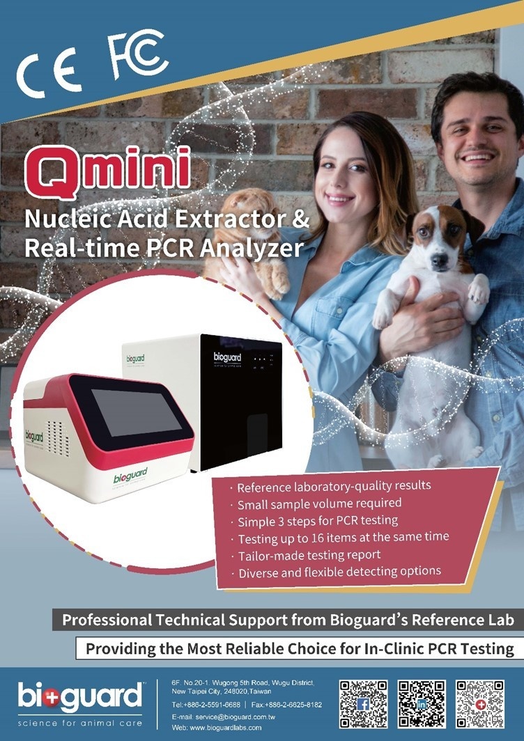
In infected cats, trichomonads are sometimes detected in the superficial mucus and mucosal crypts of colonic mucosal biopsies, accompanied by an infiltrate of lymphocytes, plasma cells, and neutrophils, as reported by Yaeger in 2005. The BIOGUARD Qmini PCR SYSTEM offers a versatile way of obtaining nucleic acid for PCR through its magnetic bead technology. In just under 10 minutes, nucleic acid samples are purified and ready for use. Additionally, the system includes a pre-made lyophilized powder PCR reaction mixer and pre-calibrated PCR protocol, resulting in PCR outcomes in 90 minutes.
4. Another method for identifying T. foetus is Fluorescence in situ hybridization (FISH).
NOTE: It is important to note that testing is most accurate in cats who have been off antibiotics for at least two weeks, as antibiotics can reduce T. foetus levels and lead to false negative results.