͏‌ ͏‌ ͏‌ ͏‌ ͏‌ ͏‌ ͏‌ ͏‌ ͏‌ ͏‌ ͏‌ ͏‌ ͏‌ ͏‌ ͏‌ ͏‌ ͏‌ ͏‌ ͏‌ ͏‌ ͏‌ ͏‌ ͏‌ ͏‌ ͏‌ ͏‌ ͏‌ ͏‌ ͏‌ ͏‌ ͏‌ ͏‌ ͏‌ ͏‌ ͏‌ ͏‌ ͏‌ ͏‌ ͏‌ ͏‌ ͏‌ ͏‌ ͏‌ ͏‌ ͏‌ ͏‌ ͏‌ ͏‌ ͏‌ ͏‌ ͏‌ ͏‌ ͏‌ ͏‌ ͏‌ ͏‌ ͏‌ ͏‌ ͏‌ ͏‌ ͏‌ ͏‌ ͏‌ ͏‌ ͏‌ ͏‌ ͏‌ ͏‌ ͏‌ ͏‌ ͏‌ ͏‌ ͏‌ ͏‌ ͏‌ ͏‌ 
Is this email not displaying correctly? View it in your browser.
Image description

Webinar: Overview of Feline Infectious Peritonitis (This webinar will be delivered in Bahasa Indonesia)

Image description

A free online class brought to you by Bioguard

Get familiar with feline infectious peritonitis. Sponsored by Bioguard Corporation and presented by Indira Putri Negari, PhD, this is the next webinar you don't want to miss it.

Access to the on-demand recording is FREE
Obtain a CERTIFICATE of attendance

REGISTER HERE

ABOUT THE WEBINAR:

Feline infectious peritonitis (FIP) is a progressive and highly fatal systemic disease of cats caused by feline coronavirus. Once a cat develops clinical FIP, the disease is usually progressive and almost always fatal. Diagnosis of FIP remains challenging and typically is the combination of signalment, clinical signs, and diagnostic aids that will help the clinician to obtain a diagnosis. In some cases, the diagnosis can be elusive and may depend on the logical exclusion of other possibilities.

ABOUT THE SPEAKER:

Dr. Indira completed her diploma degree from Faculty of Veterinary Medicine, Gadjah Mada University in Indonesia, her doctoral degree research in Biomedical Sciences and Engineering from National Central University, Taiwan. She has expertise in microbiology, immunology, and molecular biology. At present, Dr. Indira is working as a diagnostic specialist with Bioguard Corporation (Taiwan).

Image description

Monday

July 17

Image description

7 PM – 8 PM

Western lndonesian Time

Image description

Certificate of Attendance

eCertificate will be issued to the registered attendants joining the webinar for at least 50 minutes.

How to Join: Three Options:

Option 1: Watch via ZOOM

You can join us live directly via Zoom by simply registering. Please note that we will send you the link that is unique to you and should not be shared with anyone.


Option 2: Watch on our FACEBOOK Page

Follow our Facebook page and join us live during the webinar.


Option 3: Watch at your LEISURE

Registering to attend this webinar will also gain you access to the on-demand recording, which will be available 24 hours later.

SAVE YOUR SEAT

We look forward to seeing you at this event.

Happy Learning!

Want to stay up-to-date with what is going on?

Image description
Image description
Image description

Follow our Pages for the latest updates

Canine Ehrlichiosis- Symptoms and Diagnosis

Image description

Canine ehrlichiosis is a bacterial disease caused by infectious bacteria from the Ehrlichia genus. Though this disease happens everywhere, it is more common in tropical regions. With global warming, the expansion of tick habitats and the prevalence of cross-border tourism, the chances of the disease spreading to non-endemic areas have increased.


How is a dog infected with Ehrlichia?
Ehrlichiosis is a disease that develops in dogs after being bitten by an infected tick. Disease transmission can occur in as little as three to six hours after the tick attaches, therefore prompt tick removal is crucial. Ehrlichia canis (E. canis) is the most common species of the Ehrlichia genus involved in ehrlichiosis in dogs, but other species of the organism will occasionally be found.

What are the Symptoms of ehrlichiosis?

There are three phases to ehrlichiosis caused by E. canis, acute, sub-clinical and chronic. Each phase of this disease has differing or no symptoms at all.

Acute phase occurs 1 to 3 weeks after the host is bitten by the infected tick. The Ehrlichia organism is replicating in this time period and attaching to white blood cell. Symptoms for this phases of the disease include fever, lymph node swelling, limping and stiffness, reluctance to walk, reduced appetite, tiredness, coughing and breathing difficulty, and abnormal bruising and bleeding.

Dogs will generally move from the acute to the sub-clinical phase after about 1-4 weeks. In the sub-clinical phase, the organism is present but is not causing any outward signs of disease. The bacteria hide in the spleen where it can remain for months or years. Dogs that are infected sub-clinically may eliminate the organisms or may progress to the next stage, chronic ehrlichiosis.

Not all dogs ever progress from the sub-clinical phase to the chronic phase, but when they do, the symptoms become much more serious. Up to 60% of dogs infected chronically with Ehrlichia canis will have abnormal bleeding due to reduced platelet numbers.

Antibody Testing

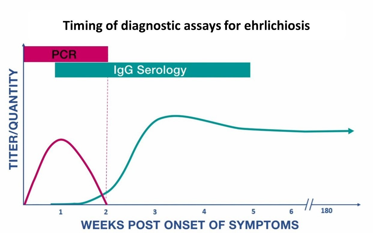
The presence of antibodies to E. canis is the basis of the serology test. Ehrlichia antibodies can be detected by serology tests, such as ELISA or rapid test. It may be difficult to diagnose infected dogs during the very early stages of infection. The immune system usually takes two to three weeks to respond to the presence of the organism and develop antibodies. In addition, antibodies against Ehrlichia species might remain elevated for many months after disease has resolved.

Real-time PCR testing

Real-time PCR testing detects the presence of Ehrlichia DNA using whole blood samples. The test is most sensitive in the first week of illness and decreases in sensitivity following the administration of appropriate antibiotics (within 48 hours). This test can also determine the species of Ehrlichia infecting dogs. Although a positive PCR result is helpful, a negative result does not rule out the diagnosis, and treatment should not be withheld due to a negative result.

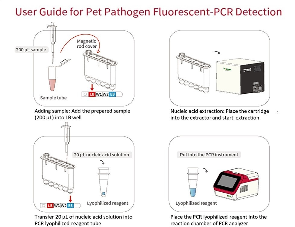
Sample collection tips for Qmini real-time PCR

Collecting more than 200 μl of blood sample, transfer to EDTA anticoagulant tube, and mix thoroughly. Then, transfer 200 μl of whole blood from EDTA tube into the sterile tube containing preservation and mix well for nucleic acid extraction.

Image description

For any direct inquiries, please contact us at: [email protected]

Diagnosis of Feline Infectious Peritonitis

Image description

Feline infectious peritonitis (FIP) is associated with a viral infection called feline coronavirus (FCoV). FIP is most common in young cats less than two years of age. Once disease develops, FIP will eventually result in death in almost every case. Reliable and rapid diagnosis of FIP is important but can be challenging.

How does FCoV cause FIP?

Most strains of FCoV are found in the gastrointestinal tract and are referred to as feline enteric coronavirus (FECV). FECV usually doesn't cause any serious issues, aside from mild diarrhea.

In some cats, mutations occur in the FECV

genome that creates a new phenotype with the ability to replicate within macrophages and cause an extreme inflammatory reaction in the tissues. The presence of highly virulent strains is referred to as the feline infectious peritonitis virus (FIPV), capable of consistently inducing FIP.

Differences between FECV and FIPV

Image description

Diagnostic tests of FIP

Image description

To diagnose FIP, a veterinarian may need to interpret several test results, as well as take into account the signalment and background of the cat.

To learn more about Rivalta testing product, click here
rapid test Ab test click here
and Qmini real-time PCR click here
Image description
Facebook
Twitter
Instagram
Facebook
Facebook

About Bioguard Corporation

The Bioguard is a company focusing on animal disease diagnostic services and products.
Our animal health diagnostic center is the first and only ISO/ IEC 17025 accredited animal disease testing laboratory in Taiwan and China.


Copyright © Bioguard Corp., All rights reserved.


Our mailing address is: [email protected]

If you want to unsubscribe, click here.