͏‌ ͏‌ ͏‌ ͏‌ ͏‌ ͏‌ ͏‌ ͏‌ ͏‌ ͏‌ ͏‌ ͏‌ ͏‌ ͏‌ ͏‌ ͏‌ ͏‌ ͏‌ ͏‌ ͏‌ ͏‌ ͏‌ ͏‌ ͏‌ ͏‌ ͏‌ ͏‌ ͏‌ ͏‌ ͏‌ ͏‌ ͏‌ ͏‌ ͏‌ ͏‌ ͏‌ ͏‌ ͏‌ ͏‌ ͏‌ ͏‌ ͏‌ ͏‌ ͏‌ ͏‌ ͏‌ ͏‌ ͏‌ ͏‌ ͏‌ ͏‌ ͏‌ ͏‌ ͏‌ ͏‌ ͏‌ ͏‌ ͏‌ ͏‌ ͏‌ ͏‌ ͏‌ ͏‌ ͏‌ ͏‌ ͏‌ ͏‌ ͏‌ ͏‌ ͏‌ ͏‌ ͏‌ ͏‌ ͏‌ ͏‌ ͏‌ 
Is this email not displaying correctly? View it in your browser.
Image description

WEBINAR SERIES

Image description

A free online class brought to you by Bioguard


Summers are here, and tick-borne diseases are much more prevalent among our pet dogs.
Learn more about the most common and frequently occurring tick-borne diseases by Dr. Yung Tsun Lo.


Access to the on-demand recording is FREE

Obtain a CERTIFICATE of attendance

REGISTER HERE
Image description

Wednesday

May 31

Image description

8 PM – 9 PM

(GMT+8)

About the Webinar:

The most important tick-borne diseases that affect dogs are Ehrlichiosis, Anaplasmosis, and Babesiosis. Transmitted by the bite of infected ticks, the severity of the disease may even lead to organ failure and death. Therefore, the healthy management plan for dogs should contain routine checks for these vector-borne diseases.

In this webinar, you will learn:
1. The cause of the three critical tick-borne diseases
2. Diagnosis of ehrlichiosis, anaplasmosis, and babesiosis in dogs
3. Treatment selections

Image description

Certificate of Attendance

eCertificate will be issued to the registered attendants joining the webinar for at least 50 minutes.

How to Join: Three Options:

Option 1: Watch via ZOOM

You can join us live directly via Zoom by simply registering. Please note that we will send you the link that is unique to you and should not be shared with anyone.


Option 2: Watch on our FACEBOOK Page

Follow our Facebook page and join us live during the webinar.


Option 3: Watch at your LEISURE

Registering to attend this webinar will also gain you access to the on-demand recording, which will be available 24 hours later.

SAVE YOUR SEAT

We look forward to seeing you at this event.

Happy Learning!

Image description

WORK-FLOW

gif-c1-drx30a-qmini-real-time-pcr-2023-05-23-062105
WATCH HERE

Contact us at [email protected] for direct inquiries

Image description

ABOUT THE WEBINAR:

Feline respiratory illness is caused by harmful pathogens such as Chlamydia felis and Mycoplasma felis. Chlamydia felis can bring about conjunctivitis, rhinitis, and pneumonia in cats. It is important to note that these bacteria can survive on surfaces such as clothing and bedding. Mycoplasma felis, on the other hand, can cause respiratory infections in cats, like pneumonia, bronchitis, and upper respiratory tract infections. It is highly contagious and can spread through contact with respiratory secretions or through fleas and other biting insects. Understanding the pathogenesis, diagnosis, and treatment of these illnesses is crucial to prevent cats from suffering from these infections.

ABOUT THE SPEAKER:

Dr. Lin got her D.V.M. degree from National Taiwan University and her Ph.D. from the College of Biological Science and Technology, National Chiao-Tung University. She is a professor in the Department of Veterinary Medicine and director of Zoonosis Research Center at National Taiwan University. In addition, she is a formal director of the Animal Disease Diagnostic Center at National Taiwan University. Her specialty includes Veterinary Clinical Microbiology, Immunology, and Animal Cancer Biology and Therapeutic Development.

REGISTER NOW

Want to stay up-to-date with what is going on?

Image description
Image description
Image description

Follow our Pages for the latest updates

Image description

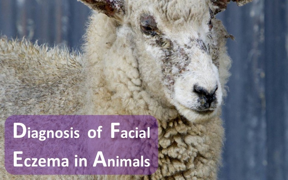
Facial eczema, also known as sporidesmin toxicosis and pithomycotoxicosis, is a disorder of grazing livestock caused by the fungus Pithomyces chartarum growing on dead plant material. It is frequently associated with perennial ryegrass pasture in New Zealand.

Clinical Findings:

Few signs are apparent until photosensitization and jaundice appear about 10–14 days after intake of the toxins. Animals frantically seek shade. Even short exposure to the sun rapidly produces the typical erythema and edema of photodermatitis in non-pigmented skin (particularly ears, eyelids, face, and lips). The animals suffer considerably, and deaths occur from one to several weeks after photodermatitis appears.

Lesions:


Histopathologic findings in the liver include degenerative changes in bile duct epithelium with both intrahepatic and extrahepatic bile ducts involved, biliary occlusion by inspissated bile and necrotic cells, hepatocellular vacuolation, particularly centrilobular hepatocytes. In a recovering liver, lesions can include the proliferation of bile ducts and periportal fibroplasia, with areas of atrophy and regeneration occurring in the liver. Characteristic liver and bile duct lesions are evident in all affected animals, whether photosensitized or not. In acute cases showing photodermatitis, livers are initially enlarged, icteric, and have a marked lobular pattern. Later, there is atrophy and marked fibrosis. The shape is distorted, and large nodules of regenerated tissue appear on the surface. In subclinical cases, livers often develop extensive areas in which the tissue is depressed and shrunken below the normal contour, which distorts and roughens the capsule. Generally, these areas are associated with fibrosis and thickening of corresponding bile ducts. The bladder mucosa commonly shows hemorrhagic or bile pigment-stained ulcerative erosions with circumscribed edema.

Diagnosis of Facial Eczema in Animals:


The clinical signs, together with characteristic liver lesions, are pathognomonic. In live animals, high hepatic enzyme activity may reflect extensive injury to the liver. Liver damage can be detected with serum chemistry changes of increased bilirubin concentration, cholesterol concentration, triacylglycerols concentration, bile acids concentration, gamma-glutamyl transaminase (GGT) activity, aspartate aminotransferase (AST) activity, glutamate dehydrogenase activity; a decrease in serum albumin concentration and increased prothrombin time can also be observed in affected animals. The diagnosis is usually based on clinical signs and the season, and a pasture spore count of Pithomyces chartarum can confirm whether the pasture is dangerous. A spore count of 100,000 or more per gram of grass is considered dangerous.

Treatment:


Supportive treatment: Treatment of the affected animals is supportive. Animals with photosensitization should be housed, provided with deep shade, and put out to graze only at night. Secondary bacterial infections should be treated as necessary. Benzimidazole fungicides on pastures: The application of benzimidazole fungicides to pastures considerably restricts the buildup of P.chartarum spores and reduces pasture toxicity.

READ MORE
Image description
Facebook
Twitter
Instagram
Facebook
Facebook

About Bioguard Corporation

The Bioguard is a company focusing on animal disease diagnostic services and products.
Our animal health diagnostic center is the first and only ISO/ IEC 17025 accredited animal disease testing laboratory in Taiwan and China.


Copyright © Bioguard Corp., All rights reserved.


Our mailing address is: [email protected]

If you want to unsubscribe, click here.